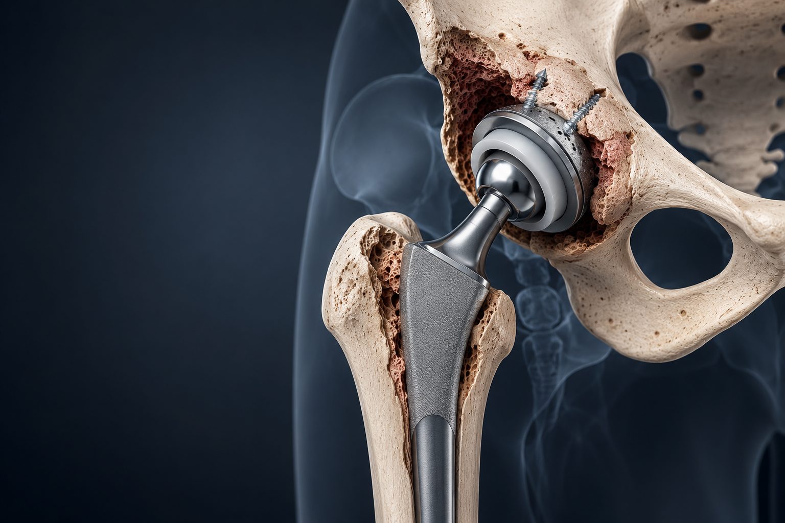
If pain has returned after your joint replacement, walking has become more difficult, you feel instability in the joint, or your doctor has recommended another operation, the first step is always to determine the cause of the problem.
Revision arthroplasty is often more complex than the original operation and requires detailed preoperative planning.
Why an implant may need a repeat operation
Modern joint replacement can restore joint function for many years, but no artificial construct lasts forever.
A repeat operation may be needed many years after the primary arthroplasty, or much sooner, if a complication develops.
The reasons can vary:
- aseptic loosening of the components
- wear of the implant's parts
- dislocation or instability
- periprosthetic fracture
- significant bone loss
- malpositioning of the components
- mechanical failure of the construct
- infectious complications
- other causes of pain and impaired function after arthroplasty
It is the underlying cause that determines the further treatment approach.
So pain after joint replacement does not automatically mean the entire implant needs to be replaced.
Your implant hurts — does that mean it needs to be replaced?
Not necessarily.
Pain after joint replacement can have many different origins. Not every cause is related to loosening of the components, and not every case requires revision surgery.
Before any decision is made, it's important to answer several questions:
- What exactly is the source of the pain?
- Are the implant components stable?
- Are they correctly positioned?
- Are there signs of infection?
- What is the condition of the bone around the implant?
- Are there other causes of impaired function?
Only after answering these questions can we determine whether surgery is needed and, if so, what its scope should be.
Aseptic loosening
Over time, one or more components of an implant can lose stable fixation to the bone.
A patient may experience pain during walking and weight-bearing, a gradual decline in function, or a sense that the joint no longer works the way it used to.
X-rays and other studies are used to assess the stability of the components and the condition of the surrounding bone.
If clinically significant loosening is confirmed, revision arthroplasty may become necessary.
Wear and bone loss
Over long-term use, an implant's components can gradually wear.
In some cases, wear particles can trigger a reaction in the surrounding tissue and lead to a gradual loss of bone — a process known as osteolysis.
This process can progress for a long time without producing noticeable symptoms.
That's why, many years after joint replacement, periodic X-ray monitoring can be important even when the implant still seems to be functioning well.
Instability and recurrent dislocation
One of the possible complications after hip replacement is dislocation.
A single episode does not always mean revision surgery is needed.
But with recurrent dislocations, the underlying cause must be identified.
It may be related to component positioning, the condition of the soft tissues, joint mechanics, or a combination of several factors.
In this situation, revision surgery should be aimed at eliminating the cause of the instability — not simply reducing the joint one more time.
Periprosthetic fracture
A fracture of the bone near an existing implant is a distinct and complex orthopedic trauma problem.
To choose the right treatment, it's important to determine:
- the location and nature of the fracture
- the stability of the implant components
- the quality of the bone
- the presence of a bone defect
- the patient's overall condition
In some situations, osteosynthesis is possible while keeping a stable implant in place.
In others, one or more components need to be replaced using revision systems.
So a periprosthetic fracture requires treating both the fracture itself and the stability of the implant at the same time.
Infection after joint replacement
Periprosthetic infection is fundamentally different from mechanical loosening.
Choosing the right approach requires confirming or ruling out an infectious process and assessing its nature.
Depending on the clinical situation, treatment may involve different strategies — from surgical debridement with retention of the components in selected cases, to single- or two-stage revision arthroplasty.
With a two-stage strategy, treatment can include:
removal of the infected components ↓ surgical debridement ↓ placement of a temporary spacer ↓ control of the infectious process ↓ repeat joint replacement
The approach is determined individually for each patient.
How revision surgery differs from the primary joint replacement
Revision arthroplasty is often technically more demanding.
The surgeon works with anatomy that has already been altered. Previous surgical approaches, scar tissue, existing components, possible bone loss, and the condition of the soft tissues all have to be taken into account.
During the operation, it may be necessary to:
- remove one or more components
- preserve any components that remain stable and suitable
- remove cement or other materials
- reconstruct bone defects
- restore a stable foundation for the new implant
- use specialized revision components
- restore the limb's length, stability, and biomechanics
Revision surgery is not simply "putting in a new implant instead of the old one." It's a reconstruction of the joint under anatomical conditions that have already changed.
Bone loss
One of the main challenges in complex revision arthroplasty is a shortage of bone.
It can result from loosening, osteolysis, infection, fracture, or previous operations.
Before revision surgery, it's important to assess:
- the location of the defect
- its size
- the quality of the remaining bone
- the options for fixing the new component
Depending on the situation, treatment may involve specialized revision components, bone grafting, augments, and other reconstructive solutions.
In the most complex cases, individual planning and patient-specific implants may be needed.
Does the entire implant need to be replaced?
Not always.
Revision arthroplasty does not necessarily mean replacing every component.
If part of the construct remains stable, correctly positioned, and compatible with the reconstruction plan, it can be preserved in appropriate cases.
In other cases, a full revision is necessary.
The scope of surgery is determined by the reason the primary arthroplasty failed and by the condition of each component individually.
01 — Reviewing the history of the primary operation
When was the joint replacement performed? Which implant was used? How did recovery go? When did the symptoms appear?
If you still have discharge summaries, operative reports, or information about the model of implant used, it's best to bring them to the consultation.
02 — Analyzing X-rays
We assess the position of the components, their stability, the condition of the surrounding bone, and any changes compared with earlier studies.
Older X-rays can be especially useful, as they allow us to see how the problem has developed over time.
03 — Additional tests
Depending on the situation, a CT scan, laboratory tests, and other diagnostic methods may be needed.
A separate priority is to rule out or confirm infection.
04 — Planning the reconstruction
We determine which components need to be replaced, which can be preserved, how large a bone defect needs to be reconstructed, and which implants may be required.
05 — Revision surgery
The planned reconstruction is carried out using the necessary revision components and methods for restoring bone support.
06 — Recovery
Your weight-bearing regimen and rehabilitation depend on the scope of the revision, the condition of the bone, and the stability of the reconstruction.
We start by establishing the cause
Pain after joint replacement is a symptom, not a diagnosis.
Before a repeat operation, it's essential to pinpoint as precisely as possible why the primary arthroplasty stopped providing the expected function.
We plan the revision before we ever reach the operating room
How complex a revision arthroplasty will be is often determined well before the operation begins.
Analyzing X-rays, CT scans, previous surgeries, and the type of implant in place allows us to prepare possible reconstructive solutions in advance.
We preserve what can be preserved
Not every component needs to be automatically removed.
If a component is stable and preserving it fits the overall surgical plan, that can be part of the chosen strategy.
We prepare for bone defects
During a revision, the surgeon must be ready not only to replace the implant, but also to reconstruct the bone support needed to fix it stably.
We combine arthroplasty with reconstructive orthopedics
Especially complex revisions can require methods that go beyond standard primary arthroplasty: bone defect reconstruction, specialized revision systems, and individualized planning.
Have you been told your implant needs to be replaced?
With revision arthroplasty, the decision can be far more complex than it was for the primary operation.
Before another procedure, it's important to understand:
- why the problem occurred
- whether revision is really necessary
- which components need to be replaced
- whether part of the implant can be preserved
- whether there is a bone defect
- whether infection has been ruled out
- what scope of reconstruction may be needed
If a repeat operation has already been proposed to you, a second opinion lets you independently assess the cause of the problem and the possible revision options before making a final decision.
How long does an implant last?
There is no universal timeframe after which an implant must be replaced.
How long it lasts depends on the type of construct, the method of fixation, the load placed on it, the condition of the bone, and many other factors.
The age of the implant on its own is not an indication for revision.
If my implant is 15–20 years old, should it be replaced as a precaution?
Not necessarily.
If it's stable, functioning well, and there's no significant wear or osteolysis, the decision to operate is usually not based on age alone.
At the same time, routine follow-up X-rays can catch changes before significant symptoms appear.
Why did my implant start hurting after several years?
There can be many reasons — from problems directly involving the components to conditions affecting the surrounding tissues or other structures.
That's exactly why diagnosing the cause of the pain is essential before any revision is considered.
Can loosening be seen on a standard X-ray?
In many cases, X-rays provide important information about the condition of the implant and the surrounding bone.
But sometimes additional imaging is needed to clarify the picture.
Can only one component be replaced?
In certain situations — yes.
If the other components are stable, correctly positioned, and compatible with the planned reconstruction, a partial revision can be considered.
What should I do if my implant has dislocated?
The first priority is to restore the correct position of the joint and assess the cause of the dislocation.
Revision is not always needed after a first episode. With recurrent instability, a more detailed workup is required.
Is a second operation more difficult than the first?
Often — yes.
During a revision, the surgeon works with altered anatomy, scar tissue, previously implanted components, and possible bone loss.
That's why preoperative planning becomes especially important.
Can an implant be reinserted after an infection?
In many clinical situations — yes.
But the approach depends on the nature of the infection, the condition of the bone and soft tissues, and the results of the earlier treatment.
Can a third or fourth operation be performed on the same joint?
The number of previous operations, on its own, doesn't determine whether another revision is possible.
What matters more is the condition of the bone and soft tissues, the stability of the construct, the presence of infection, and the options remaining for further reconstruction.
What documents should I bring to the consultation?
It's best to bring:
- your most recent X-rays
- earlier X-rays, if you still have them
- discharge summaries from the primary operation and any subsequent procedures
- information about the implant that was used
- CT and laboratory results, if these have already been done
Even old studies can be very useful, since they help us assess how the situation has changed over time.
Having problems after joint replacement?
Pain, instability, or changes seen on an X-ray don't always mean the entire implant needs to be replaced right away.
The first step is to establish the cause of the problem and assess the condition of the existing components and the surrounding bone.
During a consultation, we can review your previous treatment, X-rays, and other tests, and outline the possible options going forward.
If revision arthroplasty has already been recommended to you, the consultation can also serve as an opportunity to get a second opinion before you go ahead with another operation.
Personalized reconstructive solutions for complex orthopedic cases where a standard implant is not enough.
After a severe injury, significant bone loss, or several previous operations, the anatomy can change so much that standard off-the-shelf implants no longer offer the best solution to the reconstructive problem.
In such cases, modern CT imaging, digital 3D planning, and additive manufacturing make it possible to design a custom implant tailored to the anatomy of a specific patient.
When a standard solution may not be enough
Most orthopedic operations are successfully carried out using off-the-shelf implants in standard sizes.
But there are clinical situations in which the surgeon is not dealing with standard anatomy.
This can be caused by:
- a significant post-traumatic bone defect
- loss of part of the bone after an infection
- the consequences of multi-stage surgical treatment
- pronounced post-traumatic deformity
- anatomy altered by previous operations
- a complex defect near a joint
- the need to replace a missing bone fragment
- a situation where a standard construct doesn't match the geometry of the specific defect
In such cases, one possible solution is to design a construct for the specific patient, rather than adapting the patient to an available standard implant.
What is a custom 3D-printed implant
It is an implant whose geometry is designed to match the anatomy and specific reconstructive needs of a single patient.
It is based on computed tomography.
A digital three-dimensional model of the bone is built from the CT scan. It allows a detailed assessment of the defect, its spatial geometry, and how the anatomical structures relate to one another.
From there, the future reconstruction can be planned and the shape and parameters of the needed construct determined.
In other words, we first build a digital model of the planned reconstruction — and only then design the implant to carry it out.
01 — Computed tomography
A CT scan of the relevant segment is performed following the appropriate protocol.
The quality of this initial data is critical, since it becomes the foundation for all subsequent digital modeling.
02 — 3D reconstruction
A digital model of the bone is created from the CT scan.
It allows us to see the defect in three dimensions and assess its geometry in far more detail than a standard X-ray allows.
03 — Digital planning
We determine which anatomy needs to be restored, the position of the future construct, how it will interact with the remaining bone, and the possible fixation options.
In appropriate cases, the anatomy of the healthy opposite limb can be used as a reference for the reconstruction.
04 — Implant design
A digital model of the custom implant is created based on the surgical plan.
At this stage, the surgical and engineering sides of the process effectively merge.
05 — Manufacturing
Once the final design is approved, the construct is manufactured using the appropriate additive manufacturing technology, from a material intended for medical use.
06 — Surgery
The implant is used as part of the reconstruction that was planned in advance.
In this way, the operation doesn't begin in the operating room — much of a complex reconstruction is planned well before the surgery itself.
What individualized planning offers
The main advantage of a personalized construct isn't simply the use of 3D printing.
Its purpose is to help solve a specific anatomical problem.
Depending on the clinical situation, a custom solution can make it possible to:
- recreate the complex geometry of a missing fragment
- adapt the construct to the remaining bone
- plan the required position of the implant in advance
- restore limb length and anatomical relationships
- determine possible fixation points ahead of time
- carry out a reconstruction for which no standard implant is sufficient
3D technology is not the goal of treatment. Its value lies in the ability to carry out the reconstructive plan for a specific patient.
A custom implant isn't always the first stage
With complex post-traumatic defects, the final reconstruction can't always be performed right away.
If infection, nonviable tissue, instability, or other issues are present, a preparatory stage of treatment may be needed first.
The overall strategy may look like this:
removal of nonviable or infected tissue ↓ control of the local situation ↓ temporary stabilization or defect replacement ↓ CT scan and digital planning of the final reconstruction ↓ manufacturing of the custom implant ↓ final reconstruction
So a custom implant should be viewed as part of an overall reconstructive strategy, not as a standalone technology product.
Why the healthy limb is sometimes used
With a complex unilateral defect, part of the normal anatomy may be completely lost.
In such situations, a CT scan of the healthy opposite limb can be used to create its mirrored digital model.
This gives us a reference for what the original anatomy of the injured segment likely looked like before the injury.
The model is then adapted to the specific reconstructive task.
In effect, the healthy side can become the anatomical template for digitally restoring the injured one.
Surgeon + engineer + digital planning
Creating a custom implant is a team process.
An engineer can design a complex construct, but it's the surgeon who determines:
- what needs to be restored
- what the final anatomy should look like
- where the implant can be positioned
- how it will be inserted
- how fixation should be achieved
- how the construct fits into the overall surgical plan
That's why digital design happens through an ongoing exchange between clinical and engineering planning.
The reconstructive problem comes first — the technology second
We don't start with the question "can a 3D implant be printed?"
We first determine exactly what needs to be restored, and whether a custom construct genuinely offers an advantage over standard methods.
We plan the operation in a digital environment
The 3D model allows us to assess complex anatomy and part of the planned reconstruction before we ever reach the operating room.
We combine surgery and engineering
A custom construct isn't created separately from the operation — it's built to match a specific surgical plan.
We consider the whole course of treatment
When a multi-stage reconstruction is needed, each stage has to set up the conditions for the next one.
We don't use 3D printing where a standard solution is enough
A custom solution is justified when it helps solve a problem that would be difficult or impossible to address optimally with standard means.
Custom 3D reconstruction of a significant post-traumatic tibial defect
In complex post-traumatic cases, custom 3D planning makes it possible to move from assessing the defect to creating a personalized construct and carrying out a reconstruction that was planned in advance.
The website presents a real clinical case of reconstructing a significant tibial defect using a custom 3D-printed implant, along with the long-term functional outcome.
Can a 3D implant be made for any defect?
The technical ability to create a custom construct doesn't necessarily mean it will be the best treatment option.
The defect, the condition of the bone and soft tissues, the presence of infection, and possible standard reconstruction options all need to be assessed first.
How does a custom implant differ from a standard one?
A standard implant is mass-produced in a defined range of shapes and sizes.
A custom construct is designed for the specific anatomy and specific reconstructive needs of a single patient.
What material are these implants made from?
The material depends on the type of construct and its purpose. Custom orthopedic implants may be made from biocompatible metal materials, including titanium alloys.
The specific material is determined during the design and manufacturing of the construct.
Why is a CT scan necessary?
A CT scan is the basis for creating an accurate three-dimensional model of the bone.
A standard X-ray remains important for assessing the limb, but personalized 3D design requires spatially accurate digital data about the anatomy.
Why is a CT scan sometimes needed for both limbs?
If the defect is unilateral and part of the normal anatomy is lost, the healthy opposite side can serve as a reference.
Its digital model is mirrored and used to help reconstruct the missing anatomy.
How long does it take to manufacture a custom implant?
This is not a ready-made product taken off a shelf.
A CT scan needs to be performed, a 3D model created, a surgical plan formed, the construct designed, approved, and then manufactured.
The time required depends on the complexity of the case and the manufacturing process, and is determined individually.
Can a custom implant be placed immediately after an injury?
Not always.
With a complex injury, the initial priorities may be stabilization, removal of nonviable tissue, soft-tissue reconstruction, and infection control.
The final custom reconstruction is carried out once the right conditions for it have been established.
Can a 3D implant replace a large missing piece of bone?
In certain clinical situations, custom constructs can be part of the reconstruction of significant bone defects.
Whether this treatment is possible is determined after assessing the specific defect and the entire limb.
Is this better than bone grafting?
Not necessarily.
These are different reconstructive tools with different indications. In some cases, a biological reconstruction is the best option; in others, a custom construct or a combination of methods works better.
What should I do if I've been told there's no standard implant for my case?
The absence of a suitable standard implant doesn't automatically mean a 3D construct is needed.
In a complex situation, it's worth reviewing the CT scan, previous operations, and possible reconstructive options — including getting a second opinion on the best course of treatment.
Is a standard solution not enough for you?
If, after an injury, loss of part of the bone, or several previous operations, you've been told that a standard construct can't solve the problem, it's worth exploring the options for a custom reconstruction.
During the consultation, we'll review your test results and previous treatment and determine whether a custom 3D solution offers an advantage in your specific clinical situation.
A custom implant is not the goal of treatment. The goal is to find the reconstructive approach that best matches your specific anatomy and functional needs.